The method is based on a technique called 3D-PLI (3D Polarized Light Imaging), developed previously by the same Forschungszentrum Jülich and which is used, among other things, within the context of the important European Human Brain Project, the project financed by the European Union, which aims to describe the human brain with a level of detail never achieved before. When 3D-PLI is used, the sections of brain tissue to be examined are illuminated with polarized light. The tissue refracts the light differently depending on the polarization (i.e. in short, the angulation) given to it by the technicians, and hence – using complex technology – the spatial orientation of the nerve fibres of the brain can be calculated. This effect, called birefringence, is determined mainly by the myelin sheath, the insulating layer that surrounds nerve fibres.
However, the German and Dutch scientists went one step further, by removing a series of filters from the 3D-PLI to modify the characteristics of the polarized light (thus simplifying the diattenuation as much as possible). In this way, by combining the 3D-PLI with the diattenuation, it was possible to obtain a map of the smallest and most concealed nerve fibres – on samples taken from the brains of animals – with micrometre precision (thousandths of millimetres), distinguishing the areas of the brain with many fine nerve fibers from those with few thicker nerve fibres. It is not easy, on the other hand, to identify these types of tissues with current imaging methods.
The researchers described the results of their work in the journal Scientific Reports (of the Nature group). Thanks to this technique, it will also be possible to accumulate more knowledge on illnesses, such as multiple sclerosis or multiple system atrophy (MSA), in which damage to the myelin sheath plays an important role.
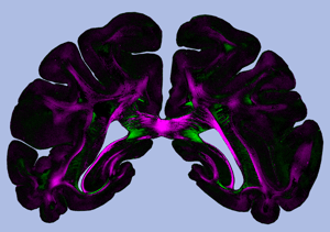
Picture: Diattenuation Imaging (DI) provides structural information about brain tissue. Copyright: Miriam Menzel et al., Scientific Reports (2019)