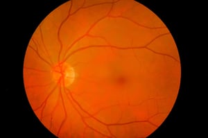
There is new hope in the treatment of serious eye diseases thanks to a new type of artificial retina developed by researchers in the Aerospace Engineering and Mechanical Engineering Department of the University of Texas, Austin campus (the retina, remember, is the very delicate nervous tissue that covers the internal part of the eye and transforms rays of light into electrical signals, which are then transported by the brain through the optic nerve). During the recent congress of the American Chemical Society in Boston (USA), the researchers explained that the new artificial retina prototype is made up of very thin layers of graphene, combined with a salt called molybdenum disulphide. The engineers also added gold, alumina and other substances to the “mix”, to create a highly flexible matrix of sensors, capable of processing the images received and sending the information to the brain.
The new material is able to take the shape of a polyhedron (like that of a 12-sided polygon or, rather, like that of a flattened soccer ball), and appears to be much more elastic than the other types of artificial retina created in the past: this is why it adapts better to the internal anatomy of the eye. Up to now prototypes have almost always been “made” of silicone, which has a relatively rigid structure, making it hard for them to replicate the natural curvature of the eye (and in addition to creating difficulties at the time of implanting the device into the eye, they also “produce” blurry or distorted images). Other attempts have also been made using organic matrices, i.e. those with a similar structure to biological tissues (as reported last year in the journal Nature Materials). However, all of these are yet to be perfected.
The path chosen by the researchers in the University of Texas is different and has achieved good results both in laboratory and animal tests: with the assistance of brain activity recordings, the researchers have in fact been able to measure the activation of the “right” areas of the visual cortex of the brain following visual stimulation. “Although this research is still in its infancy”, says Nanshu Lu, assistant professor at the University of Texas, “it is a very exciting starting point for the use of these materials to restore vision”. Should these initial good results be confirmed, it would open new doors in the future for the treatment of diseases such as retinitis pigmentosa, diabetic retinopathy, macular degeneration, traumas and other problems, which are currently very difficult to treat.